So sadly one of my emerald crabs passed away...so I ran him through the Scanning Electron Microscope at school. Morbid but really really cool.
What is a Scanning Electron Microscope you ask? Well it's exactly what its name suggests. It's a microscope which uses electrons instead of photons (light particles) to image things. Electrons being much smaller than photons of visible light provide much better resolution.
To get an image, the SEM fires electrons at the sample then detects electrons that bounce off the sample as well as those that are not.
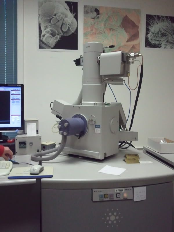
Here's what the microscope (the scope belongs to the University of Alberta's Microscopy unit) itself looks like:
Now, inorder to good good quality images on an SEM samples must first be coated in either gold or a gold-palladium mix. To do this we use this machine, a sputter coater:
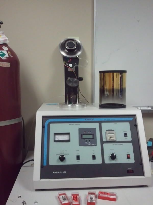
The little metal discs you see on the table are called stubs, they're essentially frag plugs for SEM specimens. You mount your specimen to your stub then you slot the stub onto disc. The disc rotates to ensure complete coverage of your specimen.
There are a few reasons we coat specimens:
1) To ensure uniform conduction of electricity.
2) To prevent charging (bright spots on the image due to differential electron absorbance/release)
Both of these are due to specimens being made up of many many different molecules/compounds etc. These will all react differently to being bombarded with electrons. The gold coating prevents solves these by providing a uniform surface.
Gold's high electron density makes it a good choice for combating these two problems.
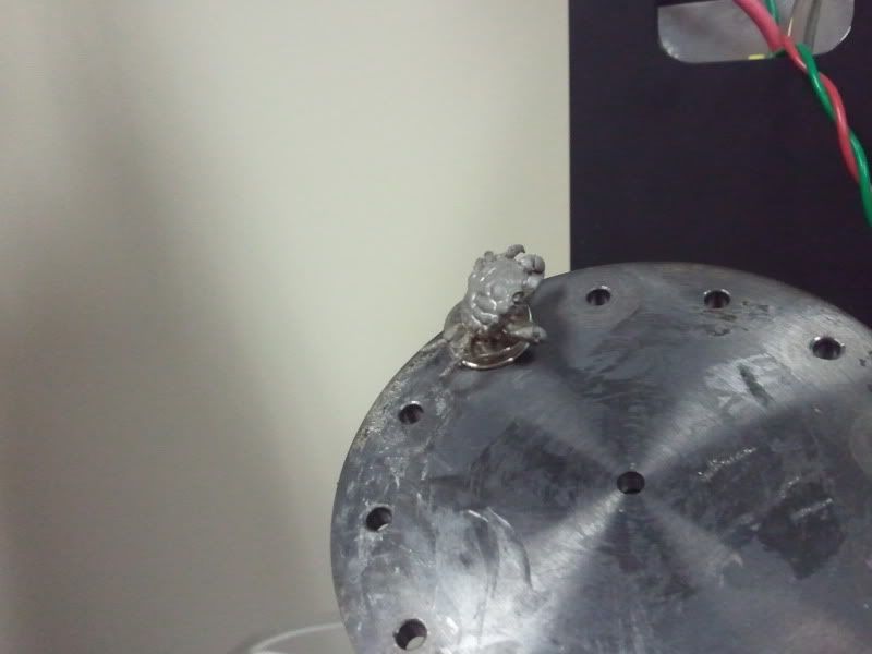
Gold-palladium coated crab:
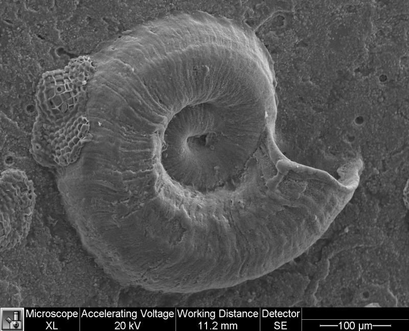
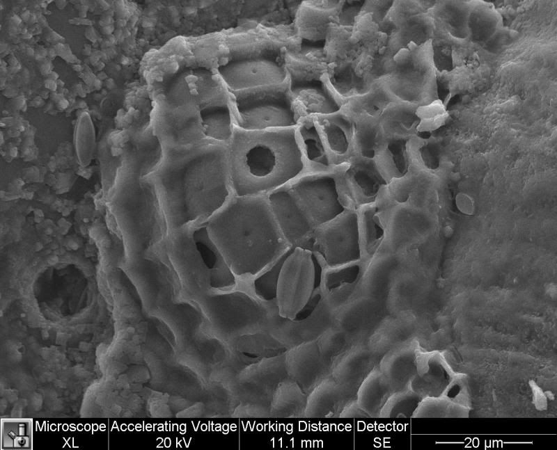
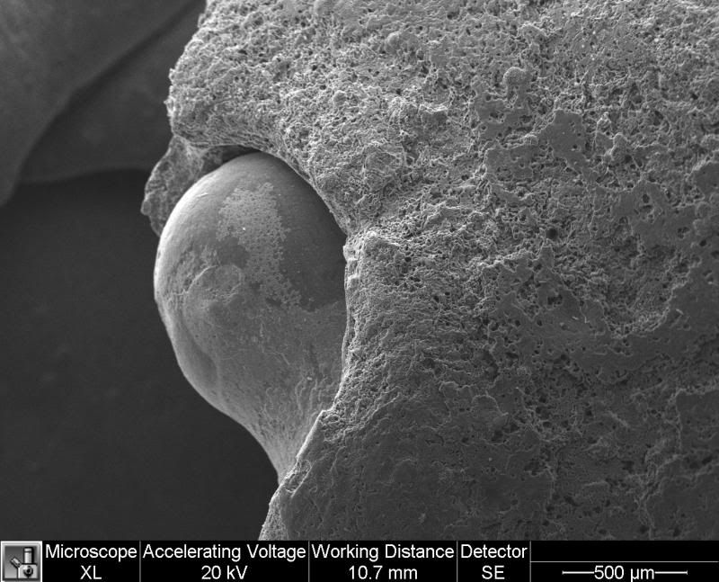
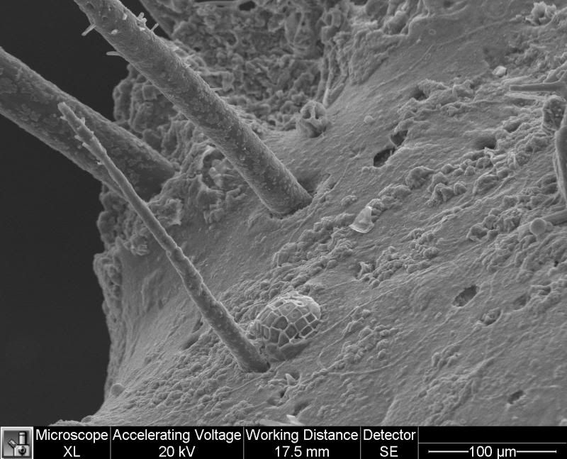
Finally, the actual images themselves. Now since the crab is so very big relative to what SEMs are normally used to look at these are actually super duper up close shots of his exoskeleton (and the things living on it!)
Eyeball:
Leg bristles:
Stayed tuned! In a few weeks I'll have histology slides of pink birdsnest!
Now I got to take these images as part of a class (Zoology 450 - Evolution of Form and Function, if anyone is interested). Actually running the SEM costs somewhere in the range of $300/hr plus some amount for the sputter coater (I'll double check the price tomorrow).
Please see wikipedia for a more through explanation of SEM:
https://en.wikipedia.org/wiki/Scanni...ion_of_the_SEM